Article

Classification, Electrophysiological Features and Therapy of Atrioventricular Nodal Reentrant Tachycardia

Register or Login to View PDF Permissions
Permissions× For commercial reprint enquiries please contact Springer Healthcare: ReprintsWarehouse@springernature.com.

For permissions and non-commercial reprint enquiries, please visit Copyright.com to start a request.

For author reprints, please email rob.barclay@radcliffe-group.com.
Average (ratings)
No ratings
Your rating

Abstract

Atrioventricular nodal reentrant tachycardia (AVNRT) should be classified as typical or atypical. The term ‘fast-slow AVNRT’ is rather misleading. Retrograde atrial activation during tachycardia should not be relied upon as a diagnostic criterion. Both typical and atypical atrioventricular nodal reentrant tachycardia are compatible with varying retrograde atrial activation patterns. Attempts at establishing the presence of a ‘lower common pathway’ are probably of no practical significance. When the diagnosis of AVNRT is established, ablation should be only directed towards the anatomic position of the slow pathway. If right septal attempts are unsuccessful, the left septal side should be tried. Ablation targeting earliest atrial activation sites during typical atrioventricular nodal reentrant tachycardia or the fast pathway in general for any kind of typical or atypical atrioventricular nodal reentrant tachycardia, are not justified. In this review we discuss current concepts about the tachycardia circuit, electrophysiologic diagnosis, and ablation of this arrhythmia.

Disclosure:The authors have no conflicts of interest to declare.

Received:

Accepted:

Correspondence Details:Dr D Katritsis, Division of Cardiology, Beth Israel Deaconess Medical Center, 185 Pilgrim Rd, Baker 4, Boston, MA, USA 02215. E: dkatrits@bidmc.harvard.edu

Copyright Statement:

The copyright in this work belongs to Radcliffe Medical Media. Only articles clearly marked with the CC BY-NC logo are published with the Creative Commons by Attribution Licence. The CC BY-NC option was not available for Radcliffe journals before 1 January 2019. Articles marked ‘Open Access’ but not marked ‘CC BY-NC’ are made freely accessible at the time of publication but are subject to standard copyright law regarding reproduction and distribution. Permission is required for reuse of this content.

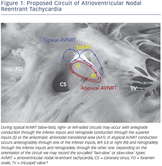
Atrioventricular nodal reentrant tachycardia (AVNRT) denotes re-entry in the area of the AV node, and represents the most common regular arrhythmia in the human.1 Although several models have been proposed to explain the mechanism of the arrhythmia in the context of the complex anatomy and the anisotropic properties of the atrioventricular (AV) node and its atrial extensions (see Figure 1),2 the actual circuit of AVNRT still remains elusive. Recent studies suggest a three-dimensional AV node with greater variability in the space constant of tissue and poor gap junction connectivity due to differential expression of connexin isoforms, that provide an explanation of dual conduction and nodal reentrant arrhythmogenesis.3,4

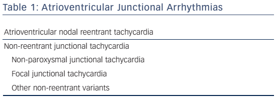
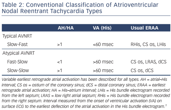
AV junctional arrhythmias are presented in Table 1. Classification schemes for AVNRT have been mainly based on the conventional concept of longitudinally dissociated dual AV nodal pathways that conduct around a central obstacle (see Table 2).

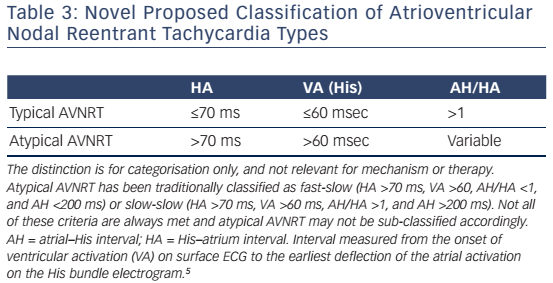
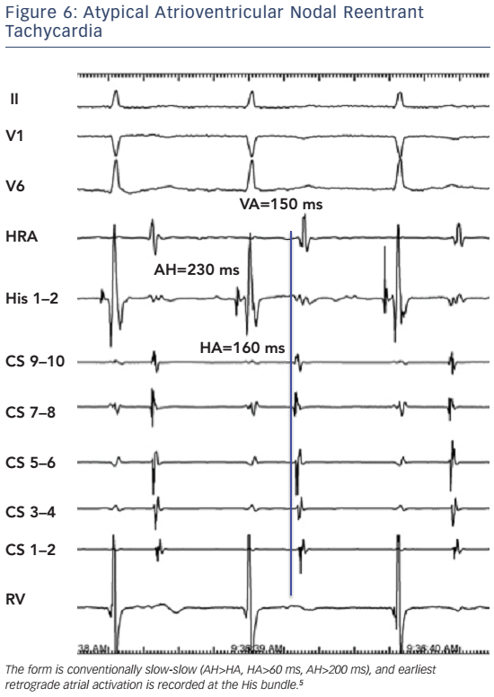
In typical slow-fast AVNRT the onset of atrial activation appears prior to, at the onset, or just after the QRS complex, thus maintaining an atrial–His/His–atrial ratio, AH/HA >1. The HA interval is usually <70 ms, measured from earliest deflection of the His bundle activation to the earliest rapid deflection of the atrial activation in the His bundle electrogram, and the VA interval, measured from the onset of ventricular activation on surface ECG to the earliest rapid deflection of the atrial activation on the His bundle electrogram, is <60 ms.1,5,6 In atypical, fast-slow form of atypical AVNRT, the retrograde atrial electrogram begins after ventricular activation with an AH/HA ratio <1. The HA interval is prolonged, ≥70 msec, and the VA interval is ≥60 msec.5,6 The atypical, slow-slow form, represents, by definition, an arrhythmia utilising two slow pathways. The AH/HA ratio is ≥1 but the HA interval is ≥70 msec, and the AH interval exceeds 200 ms.7–9

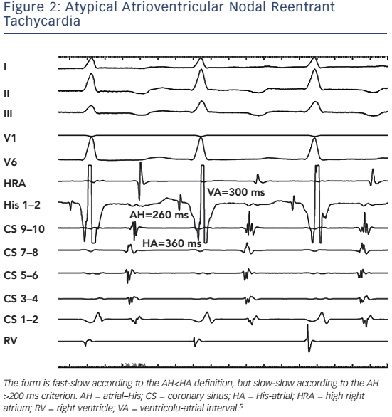
There are several inherent limitations of this classification. The distinction between fast-slow and slow-slow atypical AVNRT is often arbitrary in view of the lack of a unanimously accepted definition. In order to establish the diagnosis of a truly fast-slow form, it has been proposed that the AH interval should be less than 185 ms10 or 200 ms.6 This criterion, however, has not been adopted by other investigators.11–13 Thus, tachycardias with a relatively prolonged AH interval but an AH/ HA ratio <1 cannot be reliably classified as either fast-slow or slow-slow (see Figure 2). Furthermore, the term ‘fast-slow’ implies that the fast component of slow-fast AVNRT is the same as the fast in the fast-slow type. There is now evidence that this is not the case in patients who present with both types of tachycardia.14,15 Typical slow-fast and atypical fast-slow AVNRT appear to utilise different anatomical pathways for fast conduction. In addition, electrophysiological behaviour compatible with multiple pathways may also be seen, and in some patients, several forms of AVNRT may be inducible at electrophysiology study.

We have previously published a simplified classification scheme (see Table 2) that takes into account the shortcomings of conventional classification, and reflects evolving concepts regarding the nature of the AVNRT circuit in various forms of the arrhythmia (see Figure 1).5 AVNRT should be classified either as typical or atypical. In addition, not only the AH/HA and absolute HA intervals should be necessarily used as criteria for diagnosis of typical AVNRT. The ventriculo-atrial (VA) interval is also a practical and easily obtainable criterion, when the His bundle potential cannot be reproducibly and reliably recorded during tachycardia (see Table 3). As discussed later, retrograde atrial activation sequence or demonstration of a lower common pathway, should not be necessarily considered as reliable criteria for classification of AVNRT types.

Figure 1: Proposed Circuit of Atrioventricular Nodal Reentrant Tachycardia

Article image

Table 1: Atrioventricular Junctional Arrhythmias

Article image

Table 2: Conventional Classification of Atrioventricular Nodal Reentrant Tachycardia Types

Article image

Electrophysiological Features

Earliest Atrial Retrograde Activation

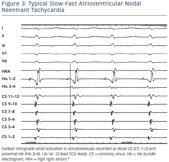
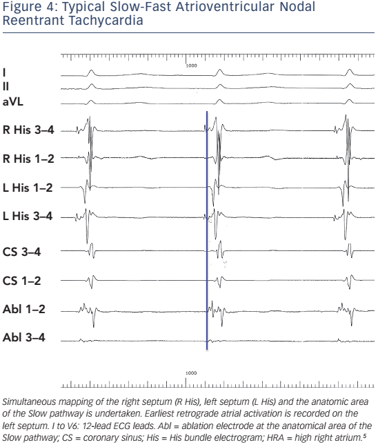
Heterogeneity of both fast and slow conduction patterns has been well described, and all forms of AVNRT may display anterior, posterior and middle retrograde activation patterns. In typical, slow-fast AVNRT, posterior or even left atrial Fast pathways may occur in ≤8 % of patients.12,13,16,17 There has also been evidence that were left septal His recordings routinely performed in patients with AVNRT, the proportion of left-sided retrograde Fast pathways might be considerably higher than previously reported.18Figure 3 and Figure 4 present typical AVNRT (slow-fast) with variable earliest retrograde atrial activation.

Figure 2: Atypical Atrioventricular Nodal Reentrant Tachycardia

Article image

Table 3: Novel Proposed Classification of Atrioventricular Nodal Reentrant Tachycardia Types

Article image

Figure 3: Typical Slow-Fast Atrioventricular Nodal Reentrant Tachycardia

Article image

Figure 4: Typical Slow-Fast Atrioventricular Nodal Reentrant Tachycardia

Article image

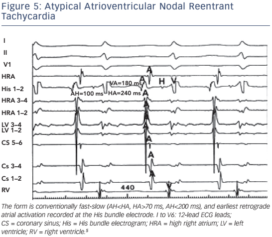
In atypical AVNRT, the earliest retrograde atrial activation is traditionally reported at the base of the triangle of Koch, near the coronary sinus ostium. Detailed mapping of retrograde atrial activation in large series of patients, however, has produced variable results. Earliest atrial activation can be well recorded at the coronary sinus ostium, the low right atrial septum, or the His bundle area.11–13,17 In certain cases of atypical AVNRT, retrograde atrial activation is even suggestive of a left lateral accessory pathway.8,9

It is obvious, therefore, that classification based on earliest atrial retrograde activation is inappropriate. Figure 5 and Figure 6 depicts fast-slow and slow-slow AVNRT, respectively, with earliest retrograde atrial activation at the His bundle electrode.

Relative AH/HA Intervals

The AH time and the relative AH/HA intervals have been proposed as a criterion for distinction between fast-slow and slow-slow AVNRT. However, both absolute and relative values may be meaningless in certain occasions. They depend on autonomic status, age, use of isoprenaline and sedatives and conduction properties of pathways involved, and may change during a single electrophysiology study. We have often noticed different AH/HA times in the same patient at similar or different tachycardia cycle lengths. Furthermore, when a His bundle electrogram cannot be recorded during tachycardia, a diagnosis based exclusively on them is impossible.19

Upper and Lower Common Pathways

Early studies have considered the possibility of additional AV nodal tissue extrinsic to the tachycardia circuit in order to explain various electrophysiologic phenomena observed during AVNRT,20 and the concepts of upper and lower common pathways have been longstanding controversies of AVNRT. The existence of an upper common pathway can now be rather easily refuted by subsequent evidence indicating that multiple atrial breakthroughs are extremely common,and retrograde activation often changes in timing and/or activation without significant alteration in tachycardia cycle, thus negating the notion of a simplistic focal atrial exit site.21–23 The perinodal transitional tissue is the route to the atrium, and in this context it may be considered as a common pathway of tissue but not a discrete site. The breakthrough is whatever leads to atrial activation via transitional tissue; thus there are many possibilities (see Figures 3–6).

Figure 5: Atypical Atrioventricular Nodal Reentrant Tachycardia

Article image

Figure 6: Atypical Atrioventricular Nodal Reentrant Tachycardia

Article image

The lower common pathway, as initially considered by Mendez and Moe, 24 has a more sound physiological basis. The notion of a lower common pathway has been utilised in order to explain phenomena of AV block without recording of a His electrogram as well as retrograde Wenckebach periodicity during AVNRT.25–28 The lower common pathway is defined as the conduction path between the distal turnaround point of the AVNRT circuit and the His bundle. The conduction time over the lower common pathway has been usually estimated by subtracting the His to atrium interval during tachycardia (measured from the onset of the His electrogram to the onset of the atrial electrogram) from that during ventricular pacing (measured from the end of the His electrogram to the onset of the atrial electrogram) at the same cycle length and considered a measurable interval in the majority of typical AVNRT cases. Initially, a lower common pathway was demonstrated in up to 75 % of 28 patients with AVNRT who were studied,20 whereas in subsequent studies with the use of para-Hisian pacing, the presence of a lower common pathway was identified in 78 % of 23 patients studied.27No evidence of a lower common pathway has been detected in typical slow-fast AVNRT.29

Table 4: Electrophysiology Techniques for the Differential Diagnosis of Narrow QRS Tachycardias

Article image

However, AV block during AVNRT without recording activation of the His bundle can also be explained by proximal intra-Hisian block.30 In up to one-third of patients with AVNRT the lower turnaround point of the circuit is within the His bundle, thus arguing against an intranodal circuit as a universal feature of AVNRT.31 Differences in the location of the lower turnaround sites of AV nodal reentry relatively to the His bundle have also been shown in experimental studies.32 Thus, block during AVNRT does not necessarily define a ‘lower common pathway’; it just defines longer refractory period below the circuit. This is often seen at the onset of very fast AVNRT, which may expose the His-Purkinje tissue to long-short periods and can lead to functional phase 3 block, having nothing to do with the reentrant circuit.

The electrophysiological proof of the existence of a lower common pathway depends on several assumptions that may not be valid, in a way that even if a lower common pathway exists, applied methodologies are unable to accurately detect and measure it.23 Furthermore, there are certain cases in the electrophysiology laboratory where an antegrade, let alone a retrograde, His bundle electrogram may not be reproducibly and reliably recorded.19 Thus, upper and lower common pathways seem to represent concepts the mechanism, relevance and practical applicability of which remain speculative.

Differential Diagnosis

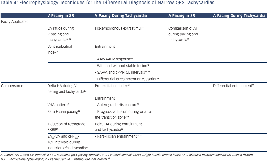
Differential diagnosis of a narrow QRS tachycardia, such as AVNRT, may be difficult.17 Although several ECG clues may assist differential diagnosis, this is usually accomplished at electrophysiology study and, most often, is between atrioventricular nodal reentrant tachycardia, atrioventricular reentrant tachycardia due to a concealed accessory pathway, and atrial tachycardia. Atrial and, mainly, ventricular pacing manoeuvres during sinus rhythm or tachycardia have been used with variable success rate. In clinical practice, these techniques cannot be applied to all cases, and multiple criteria have to be used for the differential diagnosis of narrow complex tachycardias with atypical characteristics. In Table 4 we summarise our experience with various techniques and manoeuvres for the differential diagnosis of narrow-QRS tachycardias in the electrophysiology laboratory.33–59

Ablation

Chronic administration of antiarrhythmic drugs (such as β-blockers, non-dihydropyridine calcium channel blockers, flecainide or propafenone) may be ineffective in up to 50 % of cases.1 Thus, catheter ablation is the current treatment of choice. Slow pathway ablation or modification is effective in both typical and atypical AVNRT. Usually, a combined anatomical and mapping approach is employed with ablation lesions delivered at the inferior or mid part of the triangle of Koch. 60,61 Multicomponent atrial electrograms or low amplitude potentials, although not specific for identification of slow pathway conduction, are successfully used to guide ablation at these areas. Ablation should be only directed towards the anatomic position of the slow pathway. If right septal attempts are unsuccessful, the left septal side should be tried.62,63

This approach offers a success rate of 95 %, is associated with a risk of 0.5–1 % AV block and has approximately 4 % recurrence rate. There is no mortality associated with this procedure.64,65 Advanced age is not a contraindication for slow pathway ablation.66 The preexistence of first-degree heart block may carry a higher risk for late AV block and slow pathway modification, as opposed to complete elimination, is probably preferable in this setting.67 Cryoablation may carry a lower risk of AV block, but it is negligible and this mode of therapy is associated with a significantly higher recurrence rate.67

References

  1. Katritsis DG, Camm AJ. Atrioventricular nodal reentrant tachycardia. Circulation 2010;122:831–40.
    Crossref | PubMed
  2. Katritsis DG, Becker A. The atrioventricular nodal reentrant tachycardia circuit: A proposal. Heart Rhythm 2007;4:1354–60.
    Crossref | PubMed
  3. Hucker WJ, McCain ML, Laughner JI, et al. Connexin 43 expression delineates two discrete pathways in the human atrioventricular junction. Anat Rec (Hoboken) 2008;291 : 204–15. PMCID: PMC2756604
    Crossref | PubMed
  4. Nikolaidou T, Aslanidi OV, Zhang H, Efimov IR. Structurefunction relationship in the sinus and atrioventricular nodes. Pediatr Cardiol 2012;33:890–9. PMCID: PMC3703519
    Crossref | PubMed
  5. Katritsis DG, Josephson ME. Classification of electrophysiological types of atrioventricular nodal reentrant tachycardia: a reappraisal. Europace 2013;15:1231–40.
    Crossref | PubMed
  6. Heidbüchel H, Jackman WM. Characterization of subforms of AV nodal reentrant tachycardia. Europace 2004;6:316–2.
    Crossref | PubMed
  7. Goldberger J, Brooks R, Kadish A. Physiology of “atypical” atrioventricular junctional reentrant tachycardia occurring following radiofrequency catheter modification of the atrioventricular node. Pacing Clin Electrophysiol 1992;15: 2270–82.
    Crossref | PubMed
  8. Sakabe K, Wakatsuki T, Fujinaga H, et al. Patient with atrioventricular node reentrant tachycardia with eccentric retrograde left-sided activation: treatment with radiofrequency catheter ablation. Jpn Heart J 2000;41 :227–34.
    Crossref | PubMed
  9. Vijayaraman P, Kok LC, Rhee B, Ellenbogen KA. Unusual variant of atrioventricular nodal reentrant tachycardia. Heart Rhythm 2005;2:100–2.
    Crossref | PubMed
  10. Lockwood D, Nakagawa H, Jackman WM. Electrophysiologic characteristics of atrioventriicular nodal reentrant tachycardia: implications for reentrant circuits. In Zipes DP, Jalife J. Cardiac Electrophysiology: From Cell to Bedside (Fifth edition). US: Saunders, 2009; pp.615–45.
  11. Nawata H, Yamamoto N, Hirao K, et al. Heterogeneity of anterograde fast-pathway and retrograde slow-pathway conduction patterns in patients with the fast-slow form of atrioventricular nodal reentrant tachycardia: electrophysiologic and electrocardiographic considerations. J Am Coll Cardiol 1998;32:1731–40.
    Crossref | PubMed
  12. Nam G-B, Rhee K-S, Kim J, et al. Left atrionodal connections in typical and atypical atrioventricular nodal reentrant tachycardias: activation sequence in the coronary sinus and results of radiofrequency catheter ablation. J Cardiovasc Electrophysiol 2006;17:1–7.
    Crossref | PubMed
  13. Hwang C, Martin DJ, Goodman JS, et al. Atypical atrioventricular node reciprocating tachycardia masquerading as tachycardia using a left-sided accessory pathway. J Am Coll Cardiol 1997;30:218–25.
    Crossref | PubMed
  14. Katritsis DG, Sepahpour A, Marine JE, et al. Atypical atrioventricular nodal reentrant tachycardia: prevalence, electrophysiologic characteristics, and tachycardia circuit. Europace 2015;17:1099–106.
    Crossref | PubMed
  15. Katritsis DG, Marine JE, Latchamsetty R, et al. Coexistent types of atrioventricular nodal reentrant tachycardia: implications for the tachycardia circuit. Circ Arrhythm Electrophysiol 2015;8:1189–93. PMCID: PMC4608481
    Crossref | PubMed
  16. Engelstein ED, Stein KM, Markowitz SM, Lerman BB. Posterior fast atrioventricular node pathways: implications for radiofrequency catheter ablation of atrioventricular node reentrant tachycardia. J Am Coll Cardiol 1996;27:1098–105.
    Crossref | PubMed
  17. Chen J, Anselme F, Smith TW, et al. Standard right atrial ablation is effective for atrioventricular nodal reentry with earliest activation in the coronary sinus. J Cardiovasc Electrophysiol 2004;15:2–7.
    Crossref | PubMed
  18. Katritsis DG, Ellenbogen KA, Becker AE. Atrial activation during atrioventricular nodal reentrant tachycardia: studies on retrograde fast pathway conduction. Heart Rhythm 2006;3:993–1000.
    Crossref | PubMed
  19. Katritsis DG, Josephson ME. Differential diagnosis of regular, narrow-QRS tachycardias. Heart Rhythm 2015;12:1667–76.
    Crossref | PubMed
  20. Miller JM, Rosenthal ME, Vassalo JA, Josephson ME. Atrioventricular nodal reentrant tachycardia: Studies on upper and lower “common pathways”. Circulation 1987;75:930–40.
    Crossref | PubMed
  21. McGuire MA, Lau KC, Johnson DC, et al. Patients with two types of atrioventricular junctional (AV nodal) reentrant tachycardia. Evidence that a common pathway of nodal tissue is not present above the reentrant circuit. Circulation 1991;83:1232–46.
    Crossref | PubMed
  22. Anselme F, Hook B, Monahan K, et al. Heterogeneity of retrograde fast-pathway conduction pattern in patients with atrioventricular nodal reentry tachycardia: observations by simultaneous multisite catheter mapping of Koch’s triangle. Circulation 1996;93:960–8.
    Crossref | PubMed
  23. Katritsis DG. Upper and lower common pathways in atrioventricular nodal reentrant tachycardia: refutation of a legend? Pacing Clin Electrophysiol 2007;30:1305–8.
    Crossref | PubMed
  24. Mendez C, Moe GK. Demonstration of a dual A-V nodal conduction system in the isolated rabbit heart. Circ Res 1966;19:378–93.
    Crossref | PubMed
  25. Otomo K, Okamura H, Noda T, et al. Unique electrophysiologic characteristics of atrioventricular nodal reentrant tachycardia with different ventriculoatrial block patterns: effects of slow pathway ablation and insights into the location of the reentrant circuit. Heart Rhythm 2006;3:544–54.
    Crossref | PubMed
  26. Otomo K, Nagata Y, Uno K, et al. Atypical atrioventricular nodal reentrant tachycardia with eccentric coronary sinus activation: Electrophysiological characteristics and essential effects of left-sided ablation inside the coronary sinus. Heart Rhythm 2007;4:421–32.
    Crossref | PubMed
  27. Anselme F, Poty H, Cribier A, et al. Entrainment of typical AV nodal reentrant tachycardia using para-Hisian pacing: evidence for a lower common pathway within the AV node. J Cardiovasc Electrophysiol 1999;10:655–61.
    Crossref | PubMed
  28. Kazemi B, Haghjoo M, Arya A, Sadr-Ameli MA. Spontaneous high degree atrioventricular block during AV nodal reentrant tachycardia. Europace 2006;8:421–2.
    Crossref | PubMed
  29. Heidbuchel H, Ector H, Van de Werf F. Prospective evaluation of the length of the lower common pathway in the differential diagnosis of various forms of AV nodal reentrant tachycardia. Pacing Clin Electrophysiol 1998;21 :209–16.
    Crossref | PubMed
  30. Man KC, Brinkman K, Bogun F, et al. 2:1 atrioventricular block during atrioventricular node reentrant tachycardia. J Am Coll Cardiol 1996;28:1770–4.
    Crossref | PubMed
  31. Li YG, Bender B, Bogun F, et al. Location of the lower turnaround point in typical AV nodal reentrant tachycardia: a quantitative model. J Cardiovasc Electrophysiol 2000;11 :34–40.
    Crossref | PubMed
  32. Patterson E, Scherlag BJ. Slow:fast and slow:slow AV nodal reentry in the rabbit resulting from longitudinal dissociation within the posterior AV nodal input. J Interv Card Electrophysiol 2003;8:93–102.
    PubMed
  33. Crozier I, Wafa S, Ward D, Camm J. Diagnostic value of comparison of ventriculoatrial interval during junctional tachycardia and right ventricular apical pacing. Pacing Clin Electrophysiol 1989;12:942–53.
    Crossref | PubMed
  34. Tai CT, Chen SA, Chiang CE, Chang MS. Characteristics and radiofrequency catheter ablation of septal accessory atrioventricular pathways. Pacing Clin Electrophysiol1999;22:500–11.
    Crossref | PubMed
  35. Martinez-Alday JD, Almendral J, Arenal A, et al. Identification of concealed posteroseptal Kent pathways by comparison of ventriculoatrial intervals from apical and posterobasal right ventricular sites. Circulation 1994;89:1060–7.
    Crossref | PubMed
  36. Miller JM, Rosenthal ME, Gottlieb CD, et al. Usefulness of the ΔHA interval to accurately distinguish atrioventricular nodal reentry from orthodromic septal bypass tract tachycardias. Am J Cardiol 1991;68:1037–44.
    Crossref | PubMed
  37. Owada S, Iwasa A, Sasaki S, et al. “V-H-A Pattern” as a criterion for the differential diagnosis of atypical AV nodal reentrant tachycardia from AV reciprocating tachycardia. Pacing Clin Electrophysiol 2005;28:667–74.
    Crossref | PubMed
  38. Hirao K, Otomo K, Wang X, et al. Para-Hisian pacing. A new method for differentiating retrograde conduction over an accessory AV pathway from conduction over the AV node. Circulation 1996;94:1027–35.
    Crossref | PubMed
  39. Kapa S, Henz BD, Dib C, et al. Utilization of retrograde right bundle branch block to differentiate atrioventricular nodal from accessory pathway conduction. J Cardiovasc Electrophysiol 2009;20:751–8.
    Crossref | PubMed
  40. Obeyesekere M, Gula LJ, Modi S, et al. Tachycardia induction with ventricular extrastimuli differentiates atypical atrioventricular nodal reentrant tachycardia from orthodromic reciprocating tachycardia. Heart Rhythm 2012;9:335–41.
    Crossref | PubMed
  41. Katritsis DG, Becker AE, Ellenbogen KA, et al. Effect of slow pathway ablation in atrioventricular nodal reentrant tachycardia on the electrophysiologic characteristics of the inferior atrial inputs to the human atrioventricular node. Am J Cardiol 2006;97:860–5.
    Crossref | PubMed
  42. Knight BP, Zivin A, Souza J, et al. A technique for the rapid diagnosis of atrial tachycardia in the electrophysiology laboratory. J Am Coll Cardiol 1999;33:775–81.
    Crossref | PubMed
  43. Ormaetxe JM, Almendral J, Arenal A, et al. Ventricular fusion during resetting and entrainment of orthodromic supraventricular tachycardia involving septal accessory pathways. Implications for the differential diagnosis with atrioventricular nodal reentry. Circulation 1993;88:2623–31.
    Crossref | PubMed
  44. Michaud GF, Tada H, Chough S, et al. Differentiation of atypical atrioventricular node reentrant tachycardia from orthodromic reciprocating tachycardia using a septal accessory pathway by the response to ventricular pacing. J Am Coll Cardiol 2001;38:1163–7.
    Crossref | PubMed
  45. González-Torrecilla E, Arenal A, Atienza F, et al. First postpacing interval after tachycardia entrainment with correction for atrioventricular node delay: a simple maneuver for differential diagnosis of atrioventricular nodal reentrant tachycardias versus orthodromic reciprocating tachycardias. Heart Rhythm 2006;3:674–9.
    Crossref | PubMed
  46. Bennett MT, Leong-Sit P, Gula LJ, et al. Entrainment for distinguishing atypical atrioventricular node reentrant tachycardia from atrioventricular reentrant tachycardia over septal accessory pathways with long-RP tachycardia. Circ Arrhythm Electrophysiol 2011;4:506–9.
    Crossref | PubMed
  47. Veenhuyzen GD, Coverett K, Quinn FR, et al. Single diagnostic pacing maneuver for supraventricular tachycardia. Heart Rhythm 2008;5:1152–8.
    Crossref | PubMed
  48. Segal OR, Gula LJ, Skanes AC, et al. Differential ventricular entrainment: a maneuver to differentiate AV node reentrant tachycardia from orthodromic reciprocating tachycardia. Heart Rhythm 2009;6:493–500.
    Crossref | PubMed
  49. Miles WM, Yee R, Klein GJ, et al. The preexcitation index: An aid in determining the mechanism of supraventricular tachycardia and localizing accessory pathways. Circulation 1986;74:493–500.
    Crossref | PubMed
  50. Nagashima K, Kumar S, Stevenson WG, et al. Antegrade conduction to the His bundle during right ventricular overdrive pacing distinguishes septal pathway AV reentry from atypical AV nodal reentry tachycardias. Heart Rhythm 2015;12:735–43.
    Crossref | PubMed
  51. AlMahameed ST, Buxton AE, Michaud GF. New criteria during right ventricular pacing to determine the mechanism of supraventricular tachycardia. Circ Arrhythm Electrophysiol 2010;3:578–84.
    Crossref | PubMed
  52. Dandamudi G, Mokabberi R, Assal C, et al. A novel approach to differentiating orthodromic reciprocating tachycardia from atrioventricular nodal reentrant tachycardia. Heart Rhythm 2010;7:1326–9.
    Crossref | PubMed
  53. Ho RT, Mark GE, Rhim ES, et al. Differentiating atrioventricular nodal reentrant tachycardia from atrioventricular reentrant tachycardia by DeltaHA values during entrainment from the ventricle. Heart Rhythm 2008;5:83–8.
    Crossref | PubMed
  54. Reddy VY, Jongnarangsin K, Albert CM, et al. Para-Hisian entrainment: a novel pacing maneuver to differentiate orthodromic atrioventricular reentrant tachycardia from atrioventricular nodal reentrant tachycardia. J Cardiovasc Electrophysiol. 2003;14:1321–8.
    Crossref | PubMed
  55. Pérez-Rodon J, Bazan V, Bruguera-Cortada J, et al. Entrainment from the para-Hisian region for differentiating atrioventricular node reentrant tachycardia from orthodromic atrioventricular reentrant tachycardia. Europace 2008;10: 1205–11.
    Crossref | PubMed
  56. Singh DK, Viswanathan MN, Tanel RE, et al. His overdrive pacing during supraventricular tachycardia: a novel maneuver for distinguishing atrioventricular nodal reentrant tachycardia from atrioventricular reciprocating tachycardia. Heart Rhythm 2014;11 :1327–35.
    Crossref | PubMed
  57. Man KC, Niebauer M, Daoud E, et al. Comparison of atrial-His intervals during tachycardia and atrial pacing in patients with long RP tachycardia. J Cardiovasc Electrophysiol 1995;6:700–10.
    Crossref | PubMed
  58. Sarkozy A, Richter S, Chierchia GB, et al. A novel pacing manoeuvre to diagnose atrial tachycardia. Europace 2008;10:459–66.
    Crossref | PubMed
  59. Kalbfleisch SJ, Strickberger SA, Williamson B, et al. Randomized comparison of anatomic and electrogram mapping approaches to ablation of the slow pathway of atrioventricular node reentrant tachycardia. J Am Coll Cardiol 1994;23:716–23.
    Crossref | PubMed
  60. Giazitzoglou E, Korovesis S, Kokladi M, et al. Slow-pathway ablation for atrioventricular nodal reentrant tachycardia with no risk of atrioventricular block. Hellenic J Cardiol 2010;51 : 407–12.
    PubMed
  61. Katritsis DG, Giazitzoglou E, Zografos T, et al. An approach to left septal slow pathway ablation. J Interv Card Electrophysiol 2011;30:73–79.
    Crossref | PubMed
  62. Katritsis DG, Papagiannis J. Anatomically left-sided septal slow pathway ablation in dextrocardia and situs inversus totalis. Europace 2008;10:1004–5.
    Crossref | PubMed
  63. Spector P, Reynolds MR, Calkins H, et al. Meta-analysis of ablation of atrial flutter and supraventricular tachycardia. Am J Cardiol 2009;104:671–7.
    Crossref | PubMed
  64. Bohnen M, Stevenson WG, Tedrow UB, et al. Incidence and predictors of major complications from contemporary catheter ablation to treat cardiac arrhythmias. Heart Rhythm 2011;8: 1661–6.
    Crossref | PubMed
  65. Rostock T, Risius T, Ventura R, et al. Efficacy and safety of radiofrequency catheter ablation of atrioventricular nodal reentrant tachycardia in the elderly. J Cardiovasc Electrophysiol 2005;16:608–10.
    Crossref | PubMed
  66. Li YG, Gronefeld G, Bender B, et al. Risk of development of delayed atrioventricular block after slow pathway modification in patients with atrioventricular nodal reentrant tachycardia and a pre-existing prolonged pr interval. Eur Heart J 2001;22:89–95.
    Crossref | PubMed
  67. Deisenhofer I ZB, Yin YH, Pitschner HF, et al. Cryoablation versus radiofrequency energy for the ablation of atrioventricular nodal reentrant tachycardia (the cyrano study): Results from a large multicenter prospective randomized trial. Circulation 2010;122:2239–45.
    Crossref | PubMed